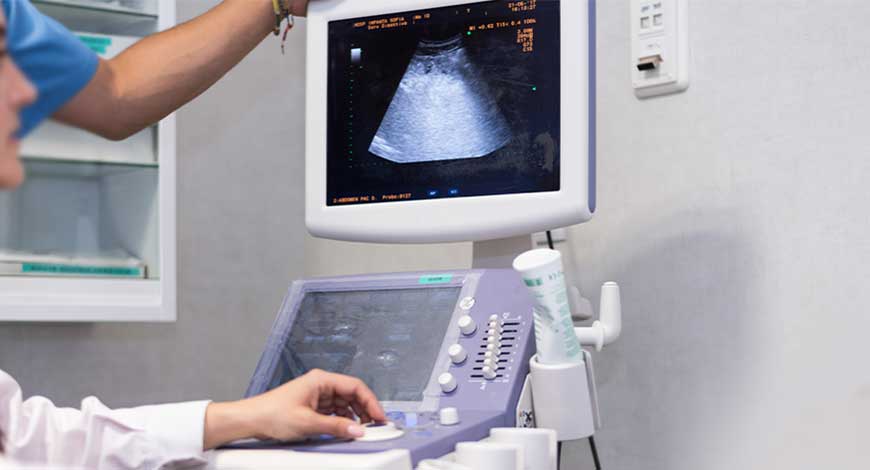
Ultrasound Equipment
Ultrasound – Leading the future of diagnostics imaging
The continuous evolution of ultrasound technology promises a future of advanced, personalized healthcare solutions. With ongoing advancements enhancing image quality and diagnostic accuracy, ultrasound continues to solidify its vital role in modern healthcare.

Ultrasound technology has become a cornerstone of modern diagnostic imaging. From detecting medical conditions to guiding life-saving procedures, ultrasound has become an indispensable healthcare tool.
Ultrasound technology, also known as sonography, has its roots in the early 20th century when physicist Paul Langevin developed sonar during World War I to detect submarines underwater. This innovation inspired its adaptation for medical purposes in the 1940s, with neurologist Dr. Karl Dussik using ultrasound to diagnose brain tumors, marking the birth of diagnostic medical ultrasound.
By the 1950s, Scottish obstetrician Ian Donald revolutionized the field by applying ultrasound to obstetrics and gynecology, earning recognition as the father of obstetric ultrasound. Machines became more refined, transitioning from cumbersome devices to compact systems. The 1960s and 1970s saw rapid advancements, expanding ultrasound’s applications to cardiology and abdominal imaging. The introduction of grayscale imaging further enhanced diagnostic precision, establishing ultrasound as a clinical mainstay.
The 1980s and 1990s ushered in a digital revolution, with portable devices and Doppler technology enabling point-of-care diagnosis and vascular imaging. In the 21st century, innovations such as high-definition imaging, 3D/4D ultrasound, and advanced Doppler techniques have transformed ultrasound into a highly versatile, accessible, and indispensable tool, shaping the future of modern diagnostics.
The journey of ultrasound is a testament to human ingenuity, merging physics, engineering, and medicine to shape the future of imaging like never before. As we continue to unlock its potential, the future of diagnostic imaging holds promises we’re only beginning to imagine.
Indian market dynamics
The Indian ultrasound systems market in 2024 is estimated at ₹1850 crore. Ongoing advancements in ultrasound technology, encompassing enhanced image resolution, 3D/4D imaging, and AI integration, have fuelled the uptake of newer ultrasound systems. This is perhaps the first year, when 40 units of super-premium machines were sold by one vendor.
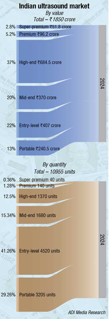
The demand shifted from mid-end segment to the high-end machines in 2024. The entry level ultrasound machines are steady at a 20-22 percent market share, and the portable at 13-14 percent.
This reflected in the numbers sold. Whereas the super-premium and the premium held their ground by combined value share, the premium machines did not gain much traction. The high-end machines saw sales of 1370 units in 2024, as compared to 660 units in 2023, while the mid-end declined from 2200 units in 2023 to 1680 units in 2024.
|
Leading players* |
|
| Tier I | GE |
| Tier II | Mindray and Samsung |
| Tier III | Philips, BPL and Sonosite |
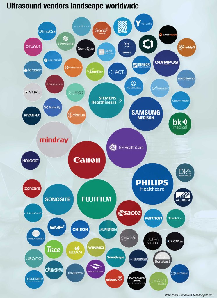
| Others | Siemens, Trivitron, Konica, Sonoscape, Esaote, Toshiba, Aloka, Fuji, Edan, Zoncare, Chison, VINNO, Wisonic, and Cura |
| *Vendors are placed in different tiers on the basis of their sales contribution to the overall revenues of the Indian ultrasound market.
ADI Media Research |
|
The entry-level machines stood their ground at 4520 units, as also the portable ones at 3205 units. Many Chinese brands made an entry in the Indian market in this segment. These include Chison, Zoncare, VINNO, and Wisonic.
In 2024, the largest share was held by diagnostic segment and is predicted to continue expanding over the coming years. Diagnostic ultrasound systems are used across various medical specialties, including radiology, cardiology, obstetrics, gynecology, internal medicine, and more. These systems are often more cost-effective compared to highly specialized ultrasound equipment. This cost advantage makes them an attractive option for healthcare institutions looking to provide comprehensive diagnostic services without substantial investments.
By application, the largest share was held by General Imaging segment and is predicted to continue expanding over the coming years. General Imaging ultrasound systems are highly versatile and can be used for a wide range of diagnostic applications. They are not limited to a specific medical specialty, making them widely applicable for various clinical needs. General Imaging ultrasound is used in multiple medical specialties, including cardiology, radiology, internal medicine, and general practice. It is employed for abdominal, pelvic, vascular, musculoskeletal, and other diagnostic purposes.
Challenges persist, notably the shortage of trained doctors in proportion to patient needs and the necessity for continuous training to optimize the usage of ultrasound systems. Addressing user expectations concerning commercial aspects and lifecycle costs remains a pivotal challenge particularly as significant machine purchases come from existing users.
Ultrasound technology – Redefining healthcare in India
 M.C. Ekambaram
Director-Marketing, UIS,
Mindray India
Ultrasound has emerged as the cornerstone of modern medicine and the device of choice. It provides cost-effective, rapid, and wide diagnostic capabilities, making it a crucial tool for improving healthcare access, particularly while managing increasing patient load. From cardiovascular conditions to cancer, ultrasound plays a vital role in early detection, diagnosis, and monitoring chronic diseases, enabling timely interventions and improved patient outcomes. Continuous innovations coupled with the integration of AI and deep learning, are enhancing ultrasound’s diagnostic accuracy at reduced scan time.
Mindray’s pioneering ultrasound solutions
Precision, early detection, accessibility & innovations in ultrasound technology are revolutionizing how we diagnose & manage diseases today.
Mindray’s ultrasound solutions are increasingly focusing on disease patterns and stages, offering a comprehensive suite of tools for:
- Early detection: Identifying conditions in their early stages for timely intervention.
- Effective evaluation: Providing detailed insights into disease severity and progression.
- Improved treatment outcomes: Guiding treatment decisions and monitoring treatment response.
Fatty liver disease serves as an excellent example of how modern ultrasound solutions address specific medical challenges. Innovative tools tailored for fatty liver include technologies such as USAT, HRI+, and LTI for early-stage assessment, HFR STE/STQ for fibrosis and cirrhosis evaluation and CEUS for focal liver lesion.
Enhancing patient care with ZST+ technology & multi-parametric solutions
At the core of these advancements lie cutting-edge technologies like ZST+ technology developed by Mindray. It is a paradigm shift from conventional beamforming to channel data-based processing which enables exceptional image quality at a very high frame rate, infinite imaging solutions & improved workflow.
Mindray’s “multi-parametric solutions” is the first of its kind in ultrasound imaging. This unique solution covers the whole body including the liver, breast, thyroid, vascular, urology, OB & GYN, and cardiology enhancing diagnostic confidence and enabling better treatment decisions. These tools support early detection and comprehensive evaluation, leading to more effective treatment outcomes.
By leveraging these advancements, modern ultrasound technology is empowering healthcare providers to deliver more precise diagnoses, optimize healthcare resource utilization & provide better healthcare for all.
Global market dynamics
The global market for ultrasound equipment is estimated at USD 9.32 billion in 2023. It is projected to grow from USD 9.99 billion in 2024 to USD 17.55 billion by 2032, growing at a CAGR of 7.3 percent from 2024 to 2032, according to Fortune Business Insights.
Recent advancements in ultrasound technology, coupled with the development of innovative equipment and improved healthcare facilities in emerging countries, have driven market players to create user-friendly devices. The rising prevalence of chronic diseases and the expanding applications of ultrasound systems are expected to propel global market growth. This demand has been complemented by innovations in point-of-care (POC) and handheld ultrasound systems, which are now widely used in primary care, anaesthesia, emergency medicine, and critical care. Recent product launches in these segments are expected to drive market growth.
The Covid-19 pandemic, had disrupted the supply chain of medical devices, negatively impacting the ultrasound market. Leading players faced challenges from distribution issues in severely affected countries such as China, India, and Brazil. Moreover, manufacturing capacities were constrained due to shortages of locally sourced raw materials and components.
In response, major market players focus on advanced systems integrated with artificial intelligence (AI). AI aims to enhance image quality, streamline interpretation, and provide real-time analysis, enabling accurate diagnosis. By addressing limitations of conventional devices—such as extended examination times, suboptimal image quality, and reliance on technicians—AI-driven ultrasound systems are poised to improve efficiency and patient outcomes.
Furthermore, the introduction of 3D/4D imaging is reshaping the ultrasound landscape, while miniaturized and portable devices are gaining traction due to their mobility and affordability. This trend toward handheld and POC systems aligns with evolving healthcare needs, fostering the growth of portable ultrasound systems.
Government and private sector funding have also provided critical support for research and development in ultrasound technology, enabling companies to develop cutting-edge devices. The growing prevalence of sports-related injuries has fuelled demand for ultrasound equipment.
Despite these advancements, challenges such as product recalls, warnings from regulatory bodies, and defective products have hindered the market’s growth. These issues tarnish brand reputations and reduce sales. Additionally, a shortage of trained technicians in countries like Australia, the UK, and Canada further hinders market expansion.
While the ultrasound equipment market faces challenges, continued innovation and targeted investments are expected to drive its growth in the coming years.
Regional insights. North America is expected to hold the industry’s largest revenue share of 42 percent by 2037. A surge in cosmetic surgeries and advancements in healthcare technologies primarily drives this growth. According to the American Society of Plastic Surgeons, cosmetic surgery procedures increased by 19 percent from 2019 to 2023, highlighting a key factor for adopting AI.
The Asia Pacific region is expected to experience substantial growth in the ultrasound, securing the second-largest market share. This growth is primarily driven by the rising prevalence of cardiovascular diseases (CVDs) and gastrointestinal disorders. This is supported by improvements in healthcare infrastructure and rising healthcare expenditures in emerging economies like India and China. These advancements address unmet medical needs and boost the demand for ultrasound systems. Furthermore, the growing adoption of retrofitted and refurbished ultrasound systems contributes to market expansion.
The increasing prevalence of chronic diseases, combined with a growing geriatric population, has driven the demand for advanced healthcare services in emerging economies. This trend has significantly contributed to the development of the ultrasound market in the Asia Pacific during the analysis period.
Point-of-care ultrasound (POCUS) is transforming healthcare in underserved areas by offering affordable, accessible, and accurate diagnostics. Expansion in POCUS availability and training local healthcare providers can bridge the imaging gap, ensuring that no child has to suffer due to a lack of vital imaging resources. Every step counts–whether it’s donating time, equipment, or expertise–together, we can bring the power of ultrasound to those in need.
Portability redefined with smarter handheld scanners
Integrating artificial intelligence (AI) into ultrasound has elevated its potential, promising enhanced diagnostic accuracy and broader applications. Notably, a ground-breaking study by the Karolinska Institute in Sweden showcased how AI models significantly improved the accuracy of ovarian cancer diagnoses. By analyzing over 17,000 ultrasound images from 3,652 patients across eight countries, these AI models achieved an accuracy rate of 86.3 percent, surpassing the 82.6 percent accuracy of human experts. This leap in diagnostic performance underscores AI’s capacity to address global shortages of skilled ultrasound examiners, particularly in detecting complex conditions like ovarian cancer.
The evolution of portable ultrasound technology has redefined diagnostics, with handheld scanners emerging as game-changers in field applications like sports injury diagnosis. These compact devices combine high-resolution imaging with AI capabilities, enabling quick and accurate assessments directly at the injury site.
Researchers at IIT-Madras have developed an indigenous portable point-of-care ultrasound (POCUS) scanner designed for sports injury diagnosis and management. This innovative device, powered by AI, allows on-field assessment of injuries, enabling immediate evaluation and treatment decisions. Developed by the Biomedical Ultrasound Imaging Lab (BUSi), the POCUS scanner targets musculoskeletal imaging and aims to bridge technological gaps in sports medicine. With safety advantages such as radiation-free imaging and sufficient resolution, it is poised to revolutionize athlete care.
Moreover, the ongoing developments in AI-powered ultrasound highlight the technology’s versatility. The Karolinska study demonstrated that AI models not only reduced misdiagnoses by 18 percent but also decreased unnecessary referrals by 63 percent, streamlining workflows in busy clinical environments.
As AI continues to reshape ultrasound technology, its applications expand into areas previously limited by resource availability and diagnostic complexity. From improving ovarian cancer detection to enabling real-time sports injury assessments, AI-driven ultrasound paves the way for smarter, faster, and more precise imaging solutions. These advancements reflect a new era in medical diagnostics, where the synergy of AI and ultrasound technology is transforming healthcare delivery across diverse domains.
The next frontier
Advancements in ultrasound technology are driving medical imaging into a new era, with 3D and 4D imaging leading the way. These cutting-edge techniques provide unprecedented detail and potentially revolutionize patient outcomes across various medical fields. From obstetrics to oncology, integrating advanced imaging systems with real-time capabilities is setting new benchmarks in diagnostics.
Building on the foundation of 3D imaging, 4D ultrasound introduces time as an additional dimension, allowing clinicians to observe dynamic processes in real time. This innovation is particularly impactful in obstetrics, enabling continuous monitoring of fetal movements and blood flow patterns.
Recent breakthroughs in high-resolution 3D ultrasound systems further redefine medical imaging precision. Systems based on 1×256 piezoelectric ring arrays have achieved remarkable accuracy, with resolutions as fine as 0.78 mm and the ability to detect lesions as small as 0.3 mm. Such precision is critical in oncology, particularly for the early detection of breast cancer. These advanced systems efficiently manage data acquisition and storage, reducing imaging complexity while delivering high-quality outputs.
The latest generation of 3D/4D volume transducers has introduced real-time imaging that enhances workflow efficiency and diagnostic accuracy. Integrating innovative tools like the Ultrasound-Derived Fat Fraction (UDFF) has further reshaped diagnostics for hepatic steatosis.
As 3D and 4D ultrasound technologies become more accessible, they are poised to significantly improve diagnostic workflows, expand applications, and improve patient outcomes.
Precision imaging in action
Shear wave elastography (SWE) represents a transformative advancement in ultrasound imaging and is poised to revolutionize diagnostic capabilities across various medical applications.
SWE transmits high-intensity ultrasound pulses at its core to generate tissue lateral shear waves. SWE encompasses various types, including point shear wave elastography (pSWE) and 2D shear wave elastography (2D-SWE). With potential applications extending to echocardiography, SWE continues to push the boundaries of ultrasound diagnostics.
One of the primary advantages of SWE is its ability to detect subtle changes in tissue stiffness, which often signify early-stage disease. For example, in liver ultrasound, SWE excels in diagnosing and staging fibrosis by quantitatively assessing liver stiffness. Similarly, breast imaging aids in differentiating benign from malignant lesions, offering an additional layer of diagnostic confidence.
Despite its many strengths, SWE is still a developing technology, with ongoing efforts to enhance its clinical reliability. The key research areas are differentiating lesion stiffness from background tissue and improving image resolution for superficial structures. Ultrasonic elastography techniques based on surface acoustic waves have emerged to address limitations in imaging superficial tissues, paving the way for broader applications and increased diagnostic accuracy.
The versatility of SWE extends beyond imaging capabilities. Its non-invasive nature, coupled with the inherent advantages of ultrasound technology—such as low cost, portability, safety, and accessibility—makes it an indispensable modality in modern healthcare.
As advancements in SWE continue to unfold, its role in medical imaging is set to expand further. From detecting liver disease and breast cancer to evaluating thyroid nodules and musculoskeletal conditions, shear wave elastography is redefining the diagnostic landscape.
Cardiac care revolutionized
Ultrasound technology is revolutionizing cardiac care by enabling precise, accessible, and non-invasive imaging. Echocardiography allows early detection of heart conditions like valve defects and blood flow abnormalities, saving lives through timely intervention. Handheld devices bring portability to the forefront, facilitating rapid bedside assessments in emergencies and remote areas.
Transesophageal echocardiography (TEE) provides real-time guidance during surgeries, ensuring precision in complex procedures. Postoperatively, ultrasound aids in monitoring valve function and detecting complications, ensuring long-term success. High-resolution imaging, 3D and 4D capabilities, and innovations like elastography enhance diagnostic accuracy, while AI automates workflows and improves consistency.
Tele-ultrasound and wireless devices expand access, allowing remote guidance and seamless mobility in critical settings. These advancements reshape cardiac care, delivering better outcomes and broader access to life-saving diagnostics and treatments.
Innovative probes and their impact
Advanced ultrasound probe technology is revolutionizing diagnostic imaging by enhancing versatility, accuracy, and safety. Multipurpose probes, capable of handling diverse diagnostic tasks, streamline workflows by eliminating frequent equipment changes. Hybrid probes, for example, combine linear and convex functionalities, allowing seamless imaging of superficial and deeper structures, making them invaluable in critical and emergency care.
Safety innovations in probe design include advanced materials and coatings that enhance durability and reduce microbial contamination. Integrated sensors for damage detection enable proactive maintenance, ensuring optimal imaging quality and patient safety. Automated disinfection systems, like UV-C and high-level disinfectors, protect probe integrity, reduce manual handling, and extend device lifespan while ensuring quick and thorough cleaning.
Building upon these advancements in ultrasound technology, the development and testing of robotics-based ultrasound probe securement systems further push the boundaries of what is possible in medical imaging. This research focuses on creating a robotics platform to improve ultrasound-based triage in emergency settings, such as military and civilian scenarios. The goal is to simplify image acquisition, which traditionally requires skilled technicians using robotics and computer vision. Four probe adapter technologies were tested for precise image capture, with two outperforming traditional methods. The next step involves integrating deep learning models for automated image capture and diagnosis, making ultrasound triage accessible and practical at the point of injury. Future efforts will focus on AI integration to enhance automated image interpretation for better medical assessment in combat casualty care.
Addressing concerns in modern ultrasound techniques
The rise of advanced ultrasound technologies like 3D and 4D imaging has revolutionized prenatal care, diagnostics, and surgical planning. However, with these advancements come concerns about the potential risks of prolonged ultrasound exposure, particularly to the fetus.
Studies confirm that, when used appropriately, 3D and 4D ultrasounds are safe, with no significant risks associated with their use. Regulatory bodies, such as the FDA, ensure the safety of these machines by controlling their energy output and recommending usage limits to prevent thermal and mechanical effects. Leading organizations like the American Institute of Ultrasound in Medicine (AIUM) and the World Health Organization (WHO) endorse these techniques when trained professionals follow established protocols.
To maximize safety, healthcare providers must follow best practices, including regular machine maintenance, continuous training, and strict adherence to scanning guidelines. Furthermore, the investment in high-quality ultrasound machines with advanced safety features, like automated exposure settings, is critical in ensuring the safety and efficacy of these imaging techniques. Regular audits and patient information about the procedure’s benefits and risks help enhance the overall safety of ultrasound imaging in clinical settings.
Therapeutic ultrasound applications are expanding. Techniques like High-Intensity Focused Ultrasound (HIFU) are employed for non-invasive surgeries, cancer treatments, and targeted drug delivery, offering less invasive treatment options for various medical conditions.
Collaborations and innovations in imaging have become pivotal in the rapid advancement of ultrasound equipment. Partnerships between academic institutions, research organizations, and medical device companies fuel the development of groundbreaking technologies that enhance the functionality and accessibility of ultrasound systems.
For example, the integration of flexible, breathable materials in ultrasound probes, such as the textile-based ultrasound imaging probe designed for long-term health monitoring, results from cross-disciplinary collaboration. By leveraging textile substrates, researchers have overcome previous challenges with air gaps and material rigidity, creating wearable, shape-conformable probes capable of continuously monitoring internal tissues. This innovation is set to revolutionize early disease detection, allowing for monitoring conditions like arteriosclerosis and dehydration through non-invasive methods.
Partnerships in ultrasound elastography are advancing the capabilities of wearable devices for continuous health monitoring. The development of bio-adhesive ultrasound elastography (BAUS-E) is a perfect example of how collaborations between medical and technological experts lead to the creation of compact, wearable systems that can track liver stiffness in real time. This innovation holds particular promise for patients in intensive care units or those recovering from organ transplants, as it enables continuous monitoring without the need for handheld probes, facilitating early detection of liver conditions such as acute liver failure.
These advancements in ultrasound imaging reflect a broader trend of leveraging partnerships and R&D to create cutting-edge solutions. Through collaborative efforts, companies and research institutions are pushing the boundaries of ultrasound technology, developing systems that are more portable and user-friendly and capable of offering real-time, continuous monitoring.
The path ahead for ultrasound technology is both innovative and transformative. Advancements in AI integration, enhanced image quality, portability, and expanding applications are solidifying ultrasound’s role as a vital tool in medical imaging and patient care across specialties. These innovations promise improved diagnostics, better treatment planning, and enhanced health outcomes worldwide, redefining medical diagnostics with smarter, more inclusive, and accessible healthcare solutions.
SECOND OPINION
Revolutionizing diagnostics – How AI-powered ultrasound is transforming healthcare in India.
Advances in ultrasound technology – Evolution of diagnostic imaging.