X-ray Equipment
From shadows to clarity – X-ray imaging in medicine and beyond
The Indian x-ray market is rapidly advancing with digital, AI, and portable technologies amid growing regulatory and cybersecurity concerns.

The legacy of Röntgen’s accidental discovery endures, as x-ray imaging continues to shape–and be shaped by–technology, clinical need, and a commitment to safer, smarter, and more accessible healthcare worldwide.
The discovery of x-rays by Wilhelm Conrad Röntgen in 1895 marks a cornerstone in the history of medicine and physics. While conducting experiments with cathode rays, Röntgen noticed a mysterious fluorescent glow emanating from a coated screen despite the cathode ray tube being encased. This led him to the realization that a previously unknown, invisible form of radiation was responsible. Capable of passing through solid objects and producing shadow-like images, he aptly named this new radiation “x-rays,” borrowing the mathematical ‘x’ symbol for the unknown.
Röntgen’s subsequent experiments rapidly demonstrated the immense potential of this discovery. His groundbreaking image of his wife’s hand, in which her bones and wedding ring were clearly visible, showcased a non-invasive view of the human body. The medical world was instantly captivated. Within a year, x-ray machines were adopted in hospitals across Europe and the United States. Physicians were suddenly able to peer inside the human body without surgery, locating broken bones, bullets, kidney stones, and foreign objects with ease. These developments laid the groundwork for the field of radiology and opened the door to future innovations such as computed tomography (CT) and fluoroscopy.
The influence of x-rays continues to ripple across medicine. By bridging the gap between basic physics and practical medical application, x-rays launched a new era of diagnostics and therapy. It allowed for more accurate diagnoses, facilitated less invasive treatments, and improved patient outcomes across the board. This legacy still echoes in every hospital and clinic today.
Indian market dynamics
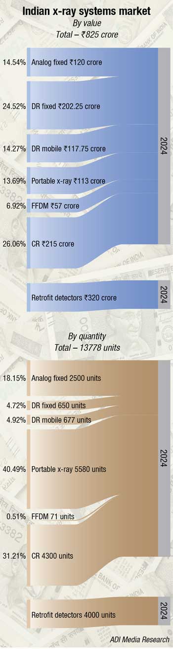
The Indian x-ray market in 2024 is estimated at ₹825 crore, with 13,778 units. Digital x-ray systems, including computed radiography and direct radiography, are rapidly replacing traditional film-based systems due to their superior image quality, faster processing, and lower radiation exposure. AI integration is accelerating, with AI-enabled solutions expected to account for up to 30 percent of radiology diagnostics by 2025.
Portable flat-panel detectors (FPDs) are gaining popularity in India due to their flexibility, high-quality imaging, and suitability for diverse healthcare settings, especially in rural and remote areas. Their ease of integration with existing systems enhances workflow efficiency. A significant impetus came in December 2024, when the Indian government introduced a 20 percent capital subsidy for domestic production of medical technology components, including FPDs. This initiative is expected to lower costs and expand accessibility, accelerating adoption across the country as demand for affordable, versatile diagnostic tools grows.
The industry is stringently regulated to ensure safety and quality, with mandatory certifications and strict standards governing the manufacturing and distribution of equipment. Growing emphasis is placed on radiation dose reduction technologies and compliance with national guidelines to protect patient health. The Atomic Energy Regulatory Board (AERB) plays a central role, overseeing licensing, type approvals, and operations, while enforcing rigorous safety codes and systematic quality assurance protocols.
The Indian government is launching a formal investigation into the increasing use of Chinese medical devices in sectors including diagnostic imaging equipment, amid heightened concerns about data security, potential cyberattacks, and national interests. The move follows a recent cybersecurity alert by the US FDA, after a “backdoor” was discovered in a Chinese-made hospital device, raising concerns about potential remote access and espionage. With China as India’s second-largest supplier after the US, officials worry that sensitive patient data collected by these devices could be misused or exploited. Rerouting of Chinese devices via third countries with which India has free-trade agreements to evade scrutiny is also of concern. If this trend persists, China could surpass the United States to become India’s largest exporter of medical devices by FY26.
Global market
As the field matures, the global x-ray equipment market is also undergoing significant transformation, driven by evolving clinical needs, technological innovation, and a rising burden of disease worldwide. Among the most profound shifts is the transition from analog to digital x-ray systems. Digital radiography is rapidly overtaking its analog predecessor due to several advantages: superior image quality, faster image processing, lower radiation exposure, and seamless data-sharing capabilities for collaborative care. This shift is being encouraged not only by performance benefits but also by regulatory pressures and contemporary reimbursement policies that prefer digital technologies. Analog and computed radiography systems are being gradually phased out in favor of more efficient alternatives.
|
Leading players* 2024 |
|
| Indian analog market | |
| Tier I | Allengers |
| Tier II | Vision, Kiran, Alerio, Medion & Epsilon; Skanray, BPL and regional fragmented brands |
| Indian CR market | |
| Tier I | Fujifilm |
| Tier II | Carestream, Konica and Agfa |
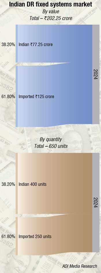
| Imported DR fixed – Full room market | |
| Imported | Fujifilm, Samsung, and Agfa. Also Philips, Siemens, Platinum, and Carestream |
| Indian | Allengers, Skanray, Alerio, BPL, and United Imaging |
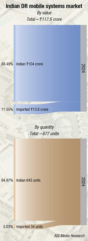
| Indian DR mobile market | |
| Imported brands | |
| Tier I | Samsung |
| Tier II | Agfa, Carestream, Shimadzu, and Mindray |
| Others | Siemens and Philips |
| Indian brands | |
| Tier I | Allengers |
| Tier II | BPL and Alerio |
| Others | Skanray and Kiran |
| Indian FFDM market | |
| Imported | Fujifilm. Also Hologics, Siemens, GE, and Planmed |
| Indian | United Imaging and Allengers |
| Indian retrofit detectors market | |
| Tier I | Konica, Fujifilm, Carestream, Loval, Agfa, LG, PZ, and CareRay |
| Tier II | Allengers, Prognosys, BPL, Skanray, Samsung, Episilon, Kiran, and Medion. |
|
*Vendors are placed in different tiers on the basis of their sales contribution to the overall revenues of the Indian x-ray market. ADI Media Research |
|
The global x-ray equipment market, valued at USD 15.6 billion in 2025, is projected to reach USD 26.23 billion by 2034, growing at a compound annual growth rate (CAGR) of 5.95 percent, according to Precedence Research. Several factors are fueling this growth. Heightened awareness regarding the importance of early disease detection is encouraging increased use of x-rays for routine and preventive diagnostics. Furthermore, the rising prevalence of chronic diseases such as cardiovascular ailments, osteoporosis, respiratory conditions, and musculoskeletal disorders–especially among aging populations–is driving up demand for diagnostic imaging services. X-rays, still among the most affordable imaging modalities, are being rapidly adopted in both primary care and specialized settings.
Indian market trends for x-ray equipment
 Sunil Rao R
Chief Technology Officer,
Skanray Technologies Limited
The digital x-ray market in India is showing promising growth at a CAGR greater than 10 percent over the next 10 years. The current value of USD 413 million is projected to reach USD 1167 million by 2033.
Growing awareness among patients and healthcare professionals is driving focus on early diagnosis and preventive care. This has increased demand for diagnostic services, with x-ray devices playing a key role. India’s ageing population and rising burden of non-communicable and infectious diseases are straining the healthcare system. The 80+ age group is projected to grow by 279 percent by 2050 (UNFPA report).
Other driving factors include the increasing accessibility of cost-effective x-ray imaging systems and solutions across a spectrum of healthcare facilities, ranging from large hospitals to smaller clinics. Technological advancements are facilitating the reduction of radiation doses in x-ray imaging, thereby enhancing patient safety and minimizing exposure.
Rising cases of accidents, falls, and sports-related injuries have increased demand for orthopaedic x-ray imaging. Routine mammography and chest x-rays for breast and lung cancer screening, respectively, are key to early cancer detection and further increase demand. The Health Card report highlights a significant projected rise in diabetes due to current lifestyles, driving the need for x-ray angiography in diagnosing and treating heart conditions. Consequently, x-ray-guided procedures, such as angiography, stent placement, and catheterisation, are becoming more prevalent, reinforcing the need for advanced x-ray systems.
Technological innovation and advancement in x-ray diagnostic and interventional devices have further accelerated the market, placing increased demand for x-ray medical devices.
Introduction of more intelligent software, AI-assisted diagnosis, reduced workflow, reduced dose levels, reduced cost of operation and teleradiology for remote diagnosis has created a high demand for devices.
There is a growing demand for portable digital x-ray systems, particularly in rural and remote areas where access to healthcare facilities is limited.
Challenges exist like device cost, lack of trained professionals, limited access in rural areas, and regulatory requirements.
The market is expected to continue its growth trajectory in the coming years, driven by the rising demand for advanced medical imaging technologies and the government’s focus on improving healthcare infrastructure.
Balancing innovation and safety
Technological advancements are continuously reshaping the capabilities of modern x-ray systems. Innovations such as three-dimensional (3D) imaging, high-resolution digital detectors, wireless portable systems, and low-dose imaging solutions are enhancing diagnostic accuracy and clinical confidence. Additionally, the integration of artificial intelligence (AI) and machine learning is streamlining imaging workflows, supporting the automation of image interpretation, and enabling quick, evidence-based decisions. These enhancements enable faster time-to-diagnosis, which can be crucial in time-sensitive clinical scenarios.
The adoption of portable and mobile x-ray units is also transforming healthcare delivery. These systems are especially useful in emergency rooms, intensive care units, home care scenarios, and remote regions lacking traditional imaging infrastructure. At the same time, techniques like photon-counting CT and spectral imaging are expanding the diagnostic breadth of x-ray systems, aiding in more precise tissue characterization and the early detection of complex diseases.
Despite this widespread innovation, several challenges persist. One major obstacle is the high initial cost of implementing digital x-ray systems, which can be particularly burdensome for facilities in lower-resource settings. Routine maintenance and regular technology upgrades can add further financial strain. Another concern is radiation exposure, especially in vulnerable populations such as children and pregnant women. These safety issues have prompted some healthcare providers to prefer modalities that do not use ionizing radiation when clinically feasible.
In response, manufacturers are advancing technologies that reduce radiation exposure without compromising image quality. Efforts include dose-reduction technologies, AI-powered imaging protocols, and comprehensive training programs centered around safer imaging practices. Such developments aim to enhance clinician confidence while ensuring patient health is safeguarded.
Outside the realm of medicine, x-ray imaging is seeing growing use in industrial and research settings. It plays a critical role in non-destructive testing, quality control, and material analysis across sectors including aerospace, automotive, and electronics. This diversification into non-medical applications is further propelling the global market and demonstrating the versatility of x-ray technology.
Expanding x-ray horizons
Pointing toward the future, the boundaries of x-ray imaging have extended into outer space. As part of the Fram2 mission, astronauts successfully captured the first-ever medical x-ray image in space, marking a pioneering milestone. The image, showing a human hand, was taken inside a pressurized spacecraft orbiting Earth at high velocity. This breakthrough demonstrates that critical diagnostic procedures, such as bone health monitoring and fracture detection, are now feasible in orbit, marking a leap in space medicine.
Executing such imaging in space conditions presents unique technical challenges. The x-ray equipment must be miniaturized, lightweight, and meet rigorous safety standards to operate in confined, zero-gravity environments. Aligning both the patient and x-ray apparatus without the benefit of gravity adds complexity, and the lack of trained radiologists onboard necessitates simplified operation protocols. Nonetheless, the image quality met clinical standards, proving the viability of in-orbit medical diagnostics.
Beyond human health, x-ray systems in space present opportunities for hardware inspection and structural integrity checks aboard spacecraft. As exploration moves toward long-duration missions, real-time, non-invasive diagnosis using x-rays will serve as a vital component of life support systems and mission safety protocols.
The transition from analog to digital imaging has profoundly reshaped the diagnostic landscape. Traditional x-ray imaging depended on film-based systems, which required chemical processing and limited real-time clinical utility. The early 2000s brought digital radiography into mainstream use, capturing x-ray images electronically and enabling immediate viewing and integrated analysis. This shift brought about immense efficiencies in clinical decision-making, allowing radiologists to adjust image contrast or zoom capabilities, enhancing diagnostic clarity while supporting remote collaboration.
Mobile radiology – Expanding access through digital innovation
 Ashu Goyal
Managing Director (Sales),
Allengers Medical Systems Ltd
Mobile radiology is revolutionizing healthcare by bringing diagnostic imaging directly to patients in ICUs, trauma care, homes, and remote regions. It ensures faster diagnosis, greater accessibility, and improved outcomes, making quality healthcare available anytime, anywhere it’s needed most.
These systems are essential in bridging the gap between patients and timely diagnostics. Ranging from manually operated low-mA units to high-power motor-driven machines, they deliver critical imaging in the most remote or sensitive environments. A key safety advantage is their low radiation dose, which makes them safe for bedside use, especially in wards such as ICUs and maternity units where vulnerable patients are present.
The evolution of digital radiography has further revolutionized this field. At the heart of this advancement lies the detector, which has replaced traditional film and CR systems. These detectors enable instant imaging, superior resolution, lower repeat rates, and reduced radiation exposure. Wireless detectors also offer unmatched flexibility, making mobile radiology faster, safer, and more efficient than ever before, particularly in time-critical environments such as trauma care or pandemic response.
In many regions, fully equipped diagnostic vans with x-ray, mammography, and ultrasound systems are extending quality care to rural populations. Additionally, MRI and CT scanners – now housed in mobile container setups-are being used in sparsely populated areas or even as backup systems in tertiary hospitals.
Handheld x-ray and ultrasound devices are making significant strides in home healthcare, and in India, portable x-ray units are actively deployed in nationwide TB screening programs.
AI-enabled mobile systems, combined with internet connectivity, allow for real-time sharing of images with experts anywhere in the world-accelerating diagnosis and improving outcomes.
Allengers has been at the forefront of this transformation with a comprehensive portfolio of mobile radiology solutions:
- MobilX DR series. Battery-powered, motor-assisted DR with up to 40 kW output, DICOM-ready, and built for mobile clinics and bedside use.
- Litex-R-DR series. Compact 6 kW & 15 kW DR systems with wireless detectors, touch controls, real-time processing, and low-dose tech.
- PORTX. Handheld x-ray system.
- X-ray vans. Fully equipped mobile units for rural and emergency deployment.
Allengers ensures imaging reaches where it matters most – quickly, safely, and digitally.
Further improvements have come through the incorporation of AI and sophisticated image processing techniques. AI algorithms now assist in detecting issues such as fractures and cancers with impressive precision, often identifying subtle signs that human observers might overlook. These algorithms can also optimize radiation dosage automatically, reducing exposure while maintaining image quality. Meanwhile, portable digital x-ray equipment–compact enough to be used in homes, ambulances, and remote clinics–makes diagnostics widely accessible, even in underserved regions.
Hospitals around the world are leveraging next-generation x-ray systems to enhance clinical services. In modern hospital settings, x-rays remain a cornerstone in diagnosing trauma, lung infections, and various internal disorders. Surgeons use real-time imaging during procedures like angioplasty for instrument guidance. Radiographers or trained assistant practitioners typically operate the imaging equipment, while radiologists interpret the images for diagnosis and treatment planning. With the deployment of advanced x-ray suites, hospitals benefit from faster scanning times, sharper imagery, and streamlined workflows, leading to more conclusive diagnoses and reduced wait times for patients.
Artificial intelligence is also elevating diagnostic accuracy in radiology. AI tools are being used successfully to interpret chest x-rays with remarkable precision, identifying early signs of infection, fractures, and chronic diseases. One such tool, Ark+, developed by researchers at Arizona State University, has demonstrated performance levels that surpass those of leading industrial software. By incorporating physician insights during its training cycle, the system can recognize complex patterns, making diagnoses more reliable and prompt.
AI doesn’t merely enhance detection; it also improves radiological workflow by flagging urgent cases, automating annotation, and reducing the burden of repetitive image review. This translates into faster reporting and clinical decision-making, which improves outcomes, particularly in high-volume hospital settings.
Technological infrastructure, such as PACS (Picture Archiving and Communication Systems) and teleradiology, is revolutionizing the delivery of radiology services globally. PACS replaces film-based image storage with cloud-based digital archives, allowing secure remote access to high-resolution scans. Seamless integration with Radiology Information Systems (RIS) and Electronic Health Records (EHRs) supports efficient data sharing and interdisciplinary collaboration. With PACS, radiologists can analyze, compare, and share medical images across hospital systems or even nationwide networks.
Teleradiology extends this capability by enabling specialists to interpret x-ray and other imaging results from almost anywhere. This is especially valuable in regions with a shortage of radiologists or limited infrastructure. High-speed communication networks and mobile technology enable radiologists to receive, assess, and report on images in real-time from smartphones or tablets. Teleradiology, often augmented by AI and machine learning, enables the prioritization of urgent cases and enhances diagnostic precision, even outside traditional clinical settings.
Green imaging
Environmental sustainability is also gaining prominence in radiology, with an emphasis on developing greener imaging solutions. Modern digital x-ray systems consume significantly less energy, reducing emissions and operating costs simultaneously. The era of hazardous, chemical-based film processing has largely ended, replaced by digital radiography that aligns with environmental regulations such as the WEEE Directive and REACH.
Dose optimization continues to be a central concern. Innovations in image reconstruction and personalized imaging protocols now facilitate high-quality diagnostic results at significantly lower radiation doses. This trend is particularly relevant for pediatric patients and routine scans where cumulative exposure must be minimized.
Detector innovations
Innovations in x-ray detector technology are reshaping the landscape of medical imaging, driving advancements toward safer and sharper diagnostic solutions. Modern detectors are now designed to minimize radiation exposure while maintaining image quality, thereby addressing both patient safety and environmental health concerns. One of the most significant developments is the creation of highly sensitive detectors that produce high-resolution images using much lower doses of radiation. This shift is especially important for frequent diagnostic procedures and for vulnerable populations like children, where cumulative radiation exposure must be carefully managed.
Technological advancements such as cascade-engineered detector configurations and the use of perovskite-based materials have contributed to reducing dark current and background noise, enhancing signal-to-noise ratios and enabling clearer imaging at significantly lower dose thresholds. Simultaneously, flat-panel detectors (FPDs) have evolved to support dual-energy imaging, allowing clinicians to distinguish between different tissue types and materials more effectively. Multi-layered detector designs now facilitate the simultaneous capture of conventional and spectral images from a single x-ray exposure, improving diagnostic accuracy while reducing patient exposure to harmful radiation.
In parallel, innovations in digital processing and image reconstruction–such as compressed sensing and deep learning algorithms–are further enhancing detector performance. These techniques enable high-quality image generation from limited data, reducing scan times and radiation doses while maintaining sharp diagnostic clarity. New detector materials and designs are also being developed to broaden the applications of imaging technologies, expanding access to advanced diagnostics even in resource-limited settings.
Together, these innovations represent a forward leap in medical imaging technology, contributing to safer, more precise, and more sustainable diagnostic practices that benefit both patients and healthcare providers alike.
Quantum materials and high-energy imaging breakthroughs
Quantum materials are further advancing the field of imaging science. Researchers at U.S. Department of Energy’s Argonne National Laboratory, in collaboration with Oak Ridge National Laboratory, Bowling Green State University, and North-western University, have created quantum shell nanoparticle-based scintillators capable of ultrafast image acquisition with exceptional spatial resolution. These materials allow for real-time imaging without blur, benefiting both scientific and clinical applications.
AI and computational advances are remapping imaging software. From 3D and 4D imaging to molecular visualization, AI enables earlier, more accurate, and personalized diagnostics. Cloud-based platforms and edge computing now support remote image processing, real-time decision support, and scalable storage. While costs and privacy concerns remain barriers, the potential for generative AI to enhance low-data image construction is opening promising avenues in imaging efficiency.
On the hardware front, innovations like cold cathode x-ray technology, precision beam shaping, and flexible liquid nanocrystal detectors are redefining portable imaging solutions. Mobile, battery-powered x-ray systems with real-time processing are making diagnostic imaging feasible in field hospitals, ambulances, and rural clinics. Some of these devices are specifically tailored for pediatric use, providing low-dose, age-appropriate examinations.
Looking ahead, the convergence of 3D imaging, quantum detectors, and sustainable design signals a new era in medical diagnostics. Volumetric 3D views enhance accuracy for orthopedic, cardiovascular, and oncology assessments. Quantum technologies are poised to redefine the standards for image quality and safety. With lightweight, modular architectures and AI-driven interpretation tools, the future of x-ray technology promises broader access, better outcomes, and greater sustainability.
From its unexpected discovery in a 19th-century lab to its cutting-edge applications in space, x-ray imaging remains one of the most dynamic and important technologies in modern science and medicine. Its trajectory reflects the remarkable power of interdisciplinary innovation, where physics, biology, engineering, and artificial intelligence converge to save lives and build a healthier, smarter future.
Second opinion