Trends
Now, ‘pencil beam’ lasers help improve brain imaging
MIT researchers say they found a way to enable faster, higher-resolution bioimaging compared to existing technology.

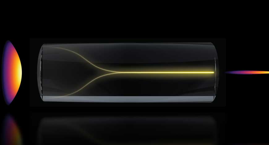
Using a “chaotic mess of laser light,” as researchers said in an April post on MIT’s website, can spontaneously self-organize into what they call a “pencil beam” under the right conditions. Using this method, researchers acquired 3D images of the human blood-brain barrier (BBB) 25 times faster than the gold-standard method. They said this maintained comparable resolution to existing technology.
The technology could show individual cells absorbing drugs in real-time, helping scientists test new drugs for neurodegenerative diseases like Alzheimer’s or ALS. This method could enable researchers to observe whether the drugs reach their targets in the brain with greater speed and resolution.
“The common belief in the field is that if you crank up the power in this type of laser, the light will inevitably become chaotic. But we proved that this is not the case. We followed the evidence, embraced the uncertainty, and found a way to let the light organize itself into a novel solution for bioimaging,” said Sixian You, assistant professor in the MIT Department of Electrical Engineering and Computer Science (EECS), a member of the Research Laboratory for Electronics, and senior author of a paper on this imaging technique.
Yu is joined on the research paper by lead author Honghao Cao, an EECS graduate student; EECS graduate students Li-Yu Yu and Kunzan Liu; postdocs Sarah Spitz, Francesca Michela Pramotton, and Federico Presutti; Zhengyu Zhang PhD ’24; Subhash Kulkarni, an assistant professor at Harvard University and the Beth Israel Deaconess Medical Center; and Roger Kamm, the Cecil and Ida Green Distinguished Professor of Biological and Mechanical Engineering at MIT.
The researchers say that they previously developed a precise fiber shaper to carefully tune laser light shining through a multimode optical fiber. They observed that, as power increased to near the point of burning, the light collapsed into a single, needle-sharp beam, rather than the expectation that the light would become more disordered and scattered.
“Disorder is intrinsic to these fibers. The light engineering you typically need to do to overcome that disorder, especially at high power, is a longstanding hassle. But with this self-organization, you can get a stable, ultrafast pencil beam without the need for custom beam-shaping components,” You said.
The researchers say they replicated the pencil beam outcome by creating two precise conditions. First, they needed the laser to enter the fiber at a perfect, zero-degree angle. Then, the power must be dialed up until the light begins to interact with the glass of the fiber itself.
In experiments, the pencil beam proved more stable and high-resolution than many similar beams. They said it was more pristine and tightly focused, while other beams often suffer from “sidelobes,” or blurry halos of light that can distort images.
The researchers then used the pencil beam in biomedical imaging of the BBB. Their technique enabled them to dynamically track how cells absorb proteins in real-time, bettering the current standard optical settings that enable the capture of one 2D section of the vasculature at a time. This requires repetition to create a fuller image.
Pencil beam imaging captured cellular-level 3D images that were higher-quality than other methods and generated those images about 25 times faster, the researchers said.
“The pharmaceutical industry is especially interested in using human-based models to screen for drugs that effectively cross the barrier, as animal models often fail to predict what happens in humans. That this new method doesn’t require the cells to have a fluorescent tag is a game-changer. For the first time, we can now visualize the time-dependent entry of drugs into the brain and even identify the rate at which specific cell types internalize the drug,” said Kamm.
Next, the researchers hope to better understand their method’s fundamental physics and the mechanisms behind its self-organization. They also hope to apply the technique to other scenarios, like imaging neurons in the brain, ahead of potential commercialization.
“You’s group realized this beam that concentrates energy in time and space could be valuable for microscopy techniques that depend on the intensity of the light that illuminates the sample. They demonstrated just that and found advantages over ordinary laser beams for imaging. It will be scientifically interesting to fully understand the creation of the new pencil beams, which could find use in a variety of imaging applications,” said Frank Wise, the Samuel B. Eckert Professor of Engineering Emeritus at Cornell University, who was not involved with this work. Medical Design & Outsourcing