MB Stories
Mammography’s next chapter – Smarter imaging, scalable screening, equitable access
Personalized, AI-enabled mammography is redefining breast cancer detection, expanding access, and transforming screening workflows.

Breast cancer remains one of the most pressing global health challenges, underscoring the need for urgent, effective, and equitable screening. Mammography has underpinned early detection strategies for decades, improving survival by identifying disease at more treatable stages. Yet access to quality screening remains uneven, particularly across low- and middle-income countries where infrastructure gaps, affordability constraints, and acute radiologist shortages persist. Large populations continue to miss timely, routine examinations, and late-stage diagnosis still carries a heavy clinical and economic burden for health systems.
In response, breast imaging is undergoing a rapid, innovation-led transformation worldwide. Researchers, clinicians, and technology developers are building solutions that are more affordable, robust, and adaptable to diverse care settings. AI, advanced imaging platforms, and sophisticated data analytics are enhancing the sensitivity and specificity of early detection, often by identifying subtle radiologic signatures of disease. These tools increasingly complement conventional mammography and are being deployed in rural clinics, community health centres, and large-scale public screening initiatives.
Legacy imaging technologies are being systematically upgraded. Digital systems, three-dimensional visualization, contrast-enhanced techniques, and data-driven diagnostic support are sharpening clinical accuracy and accelerating decision-making. AI is woven into radiology workflows to flag suspicious patterns, reduce inter-reader variability, and improve overall screening performance.
Globally, early-detection efforts are being reinforced through awareness initiatives, organized screening programs, and broader preventive health strategies. Governments, health systems, and provider networks are prioritizing population-based screening to reduce breast cancer mortality. Funding is flowing into next-generation imaging systems, mobile screening units, and integrated digital platforms that extend reach into underserved geographies.
Breast cancer detection is thus evolving from basic imaging toward integrated, technology-enabled ecosystems. High-performance hardware, AI-driven analysis, and seamless digital connectivity are coalescing into the next generation of mammography. The objective is clear: high-precision detection, scalable screening capacity, and truly inclusive access that enables earlier diagnosis and improved survival.
Democratizing breast cancer screening – Indian innovation for a new era in mammography
 Ashu Goyal
Managing Director (Sales),
Allengers Medical Systems Ltd
Breast cancer is the most common and fatal cancer among Indian women. The growing burden of the disease has driven a shift in healthcare priorities from treating advanced-stage cases to promoting early detection through organized screening programs. This transition calls for rethinking how mammography technology is designed, deployed, and delivered across India, especially with mobility and accessibility becoming central to the screening ecosystem.
Bridging the accessibility gap
Historically, mammography services in India were concentrated in metropolitan hospitals, limiting access for women in smaller cities, towns, and rural areas. Today, Indian innovation is helping bridge this gap by developing technologies that can reach women where they live and work.
A key development is modular portability. Modern mammography systems are being designed to be compact, durable, and suitable for installation in mobile screening vans. These mobile units enable healthcare providers to conduct screening camps in semi-urban and rural regions, ensuring that quality diagnostic services are no longer restricted to major healthcare centers.
Another important shift is decentralized deployment. Mammography technology is gradually moving beyond tertiary hospitals to diagnostic centers, community health facilities, and workplace health programs. This broader distribution improves screening participation and encourages early detection.
Precision through localized intelligence
While accessibility is essential, maintaining diagnostic accuracy remains critical. Advanced software innovations are enhancing the clinical value of mammography systems.
AI-driven clinical support is increasingly integrated to assist radiologists by acting as a second pair of eyes. AI tools help identify suspicious lesions, highlight abnormalities, and prioritize high-risk cases for closer evaluation.
Additionally, tailored imaging algorithms are being developed to address dense breast tissue, commonly observed among Indian women. Optimized image processing improves lesion visibility and supports reliable diagnosis.
Scaling sustainable solutions
For mammography screening to expand nationwide, sustainability must also be addressed.
Indigenous R&D and manufacturing play an important role in reducing dependence on imports while ensuring faster technical support and service availability. Local innovation helps make advanced systems more affordable for healthcare providers.
We at Allengers are contributing to this effort by developing reliable, cost-effective mammography solutions designed for diverse healthcare settings, including mobile screening initiatives, helping expand access to quality breast cancer screening.
The market
Technology innovation is a central growth catalyst. Digital mammography, three-dimensional tomosynthesis, and AI-enabled image analysis are transforming traditional systems into high-precision diagnostic platforms. These technologies enhance the detection of faint abnormalities, reduce reading times, and support higher patient volumes. AI brings additional value through automated image interpretation, decision support, and workflow optimization. By mining large imaging datasets, AI tools can refine malignancy risk assessment, reduce false positives and negatives, and enable more tailored screening pathways.
Supportive policy environments are reinforcing the trend. Governments are investing in national screening programs, expanding reimbursement for breast imaging, and funding infrastructure upgrades. Public awareness campaigns, combined with capital allocations for diagnostic equipment and digital platforms, are increasing demand for cutting-edge mammography systems. Underserved regions are increasingly targeted through mobile units, cloud-enabled platforms, and teleradiology, guided by national mandates for early detection.
At the same time, market constraints remain significant. The high acquisition and lifecycle costs of digital and 3D mammography systems limit adoption by smaller facilities and resource-constrained providers. Many developing markets still rely on analog equipment because of lower upfront costs, despite trade-offs in image quality and workflow efficiency. Workforce limitations–particularly shortages of radiologists and trained technologists–constrain utilization of advanced systems. Regulatory complexity, delayed reimbursement decisions, and macroeconomic headwinds can slow procurement cycles and capacity expansion.
Industry partnerships are emerging as important enablers. Medical technology manufacturers are collaborating with AI specialists to embed analytics directly into mammography systems. Hospital–industry–research alliances are accelerating technology validation and the generation of clinical evidence for new screening approaches. Government, NGO, and device-maker collaborations are expanding screening coverage in remote and underserved regions. These strategic initiatives are catalysing innovation, addressing access barriers, and supporting the emergence of integrated detection ecosystems that underpin sustained market growth.
In India, several drivers are converging – Nationwide and regional awareness campaigns, expansion of diagnostic infrastructure into Tier-II and Tier-III cities, and a growing role for private healthcare providers. Government cancer-control programmes and subsidized screening initiatives are adding impetus to market growth. Nonetheless, infrastructure gaps, affordability challenges, and regional inequities remain, particularly in rural and remote areas, underscoring the need for cost-effective and scalable solutions.
Competitive edge sharpens
Competition across the mammography segment is intensifying, with technology differentiation, digital capabilities, and ecosystem partnerships emerging as key battlegrounds. Leading manufacturers are focusing on AI-integrated platforms, tomosynthesis, and automated analysis tools that offer higher diagnostic precision and more efficient workflows.
Substantial R&D investments are directed toward next-generation systems that deliver higher image quality, lower radiation dose, faster acquisition, and improved ergonomics. Collaborations with telehealth providers, AI firms, and large hospital networks are enabling faster deployment of advanced solutions and extending reach into non-metro and emerging markets.
To address cost pressures and market access barriers, manufacturers are localizing production, optimizing supply chains, and developing portfolio tiers suited to different price and performance points. Competitive dynamics are gradually shifting from volume and price-based competition to innovation-led, digitally enabled, and sustainability-conscious strategies that favour companies with strong technology and partnership capabilities.
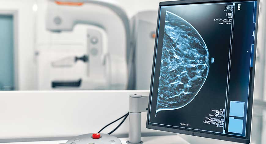
From 2D mammography to 3D tomosynthesis
Over recent decades, breast imaging has advanced from film-based mammography to digital platforms and now to three-dimensional tomosynthesis, each step addressing specific diagnostic limitations.
Conventional film-based mammography captured x-ray images on radiographic film for manual interpretation. While instrumental in the early detection of breast cancer, analog systems were constrained by limited dynamic range, suboptimal contrast resolution, and operational challenges related to image storage, retrieval, and sharing.
Digital mammography represented a major inflection point. Electronic detectors capture images that are processed and stored digitally, enabling post-processing, magnification, contrast adjustment, and integration with PACS and hospital information systems. This shift significantly improved image quality, facilitated side-by-side comparison with prior studies, and enhanced workflow efficiency.
However, two-dimensional imaging, even in digital form, is inherently limited by tissue overlap. The breast is a three-dimensional structure, and the superimposition of tissues in a flat 2D projection can obscure lesions or create artefacts that mimic pathology. This reduces sensitivity and may increase false-positive rates, requiring additional imaging or interventions.
Digital breast tomosynthesis (DBT) was introduced to mitigate these limitations. Tomosynthesis acquires multiple low-dose images from different angles during a single breast compression and reconstructs them into thin slices across the breast volume. Radiologists can scroll through these slices to evaluate tissue layer by layer, reducing the impact of tissue overlap.
Clinical evidence demonstrates that DBT increases cancer detection, particularly for invasive cancers, while reducing recall rates for additional imaging. Patients benefit from fewer unnecessary callbacks and more definitive diagnostic information.
Tomosynthesis is especially advantageous in women with dense breasts, where conventional mammography struggles with reduced sensitivity. Many contemporary systems offer combined 2D and reconstructed 3D imaging within a single examination, integrating both modalities into routine practice.
The progression from analog to digital and now to 3D tomosynthesis exemplifies breast imaging’s continuous drive toward higher precision, greater sensitivity, and more individualized care, laying a strong foundation for further integration with AI and advanced analytics.
Next-generation detector technology
Detector and hardware innovation is central to the next wave of mammography performance gains. Detecting small, low-contrast lesions in complex breast tissue requires imaging systems capable of very high sensitivity, excellent spatial resolution, and efficient dose utilization.
New detector architectures incorporate advanced scintillator materials, highly sensitive photodiodes, and improved readout electronics to capture more information from each x-ray exposure. This combination enhances contrast resolution and sharpness while supporting lower radiation doses.
Photon-counting detectors mark a particularly important advance. Unlike conventional energy-integrating detectors, photon-counting systems detect and individually count incoming x-ray photons, often with energy discrimination. This enables better noise performance, higher contrast-to-noise ratios, and the potential for spectral imaging, where different tissue types can be better differentiated based on energy-dependent attenuation.
High-resolution digital arrays, optimized scintillation coupling, and sophisticated signal processing algorithms produce images with wide dynamic range and fine structural detail. These capabilities improve visualization of microcalcifications and subtle architectural distortion, which are critical markers in early breast cancer.
As these detector technologies mature, they are expected to underpin the next generation of mammography systems, offering improved diagnostic performance while maintaining or reducing dose, and supporting new imaging paradigms such as spectral and quantitative mammography.
AI transforming mammography interpretation
AI is now a central component of the evolving mammography ecosystem, particularly in image interpretation and decision support. Deep learning algorithms, trained on large, curated datasets of mammograms and annotated outcomes, can recognize complex imaging patterns associated with benign and malignant lesions.
These systems analyse multi-view mammograms and, increasingly, integrate information from other modalities such as tomosynthesis, ultrasound, or MRI. AI outputs typically include region-of-interest highlighting, malignancy probability scores, and structured descriptors aligned with reporting standards.
In clinical practice, AI is being deployed as a concurrent or second reader, supporting radiologists in detecting lesions and standardising assessments. Early studies indicate that AI can increase sensitivity, reduce missed cancers, and potentially lower false-positive rates when used appropriately within a human–machine workflow.
AI also has a growing role in screening operations. In high-volume programmes, algorithms can pre-screen exams, identify high-risk cases for prioritized review, and streamline the reading queue. This is particularly valuable in resource-limited contexts where radiologist capacity is constrained.
Emerging AI approaches leverage multimodal and longitudinal data. Models trained on imaging plus clinical parameters such as age, family history, hormonal factors, and prior imaging can generate more refined, patient-specific risk estimates. Self-supervised learning and large foundation models further expand AI’s capabilities by learning from massive datasets with limited manual labelling.
Importantly, AI in mammography is positioned as an augmentation tool rather than a replacement for radiologists. When integrated thoughtfully, it can enhance accuracy, reduce fatigue, and support more consistent, evidence-based decision-making.
Mammograms as multi-disease diagnostic platforms
The diagnostic potential of mammography is expanding beyond breast cancer. High-quality mammograms, when coupled with advanced analytics, can provide insight into other aspects of systemic health.
One of the most studied examples is breast arterial calcification (BAC), which is often visible on standard mammograms. These calcifications are increasingly recognized as correlates of cardiovascular risk. AI and quantitative image analysis can automatically detect and measure BAC, creating an opportunity to integrate cardiovascular risk assessment into routine breast screening without additional imaging or radiation.
Beyond vascular markers, computational imaging techniques can mine mammograms for a wider array of imaging biomarkers linked to metabolic status, endocrine factors, and tissue microenvironment. Large-scale analyses of imaging phenotypes may reveal associations with non-breast malignancies, metabolic disorders, or other systemic conditions.
This evolution positions mammography as a multi-purpose, data-rich diagnostic platform. By extracting additional clinically relevant information from an existing exam, health systems can maximize the value of screening encounters, strengthen preventive care, and open new pathways for risk stratification and early intervention.
Clinical workflow transformation
AI is also reshaping how radiology departments organize and deliver services. Mammography workflows, traditionally labour-intensive and dependent on manual triage and double reading, are being reconfigured around automation and intelligent case management.
AI solutions can conduct a preliminary pass through incoming studies, flagging those with high suspicion for cancer, assigning risk categories, and routing cases accordingly. Low-risk studies may be expedited, while complex or ambiguous cases receive focused attention from experienced readers.
Evidence from early deployments suggests that AI can reduce the number of cases requiring human double reading while maintaining comparable detection and recall performance. This has important implications for screening programmes facing staffing constraints, particularly in public sector and rural settings.
Visualization tools embedded in AI platforms highlight suspected lesions, provide risk scores, and offer structured prompts, helping radiologists review studies more efficiently. This can reduce interpretation time and variability, while supporting adherence to reporting standards.
AI-driven risk models also enable prospective planning. By using imaging-based and clinical risk scores, programmes can assign women to different screening pathways–such as annual versus biennial screening, supplemental imaging, or targeted follow-up–optimising resource allocation and improving outcomes.
The net effect is a shift toward human–AI collaboration, where AI handles high-volume, repetitive tasks and radiologists focus on complex interpretation, multidisciplinary discussions, and patient communication.
Real-world AI screening
Across geographies, hospitals and diagnostic centres are moving from pilot projects to routine clinical deployment of AI-enabled mammography. Modern platforms combine advanced detectors, automated exposure control, and high-performance reconstruction with integrated AI analysis.
These systems generate high-quality images at low radiation dose, including in women with dense breast tissue–a long-standing challenge in screening. AI modules can automatically identify suspicious regions, calculate risk scores, and provide structured outputs that integrate into reporting systems.
As these platforms become more widely available, they are spreading beyond tertiary centres to regional and mid-size facilities. Combined with cloud connectivity and teleradiology, this is enabling more uniform access to advanced breast imaging across urban and non-urban areas.
Operationally, real-world deployments have demonstrated improvements in reading efficiency, more consistent reporting, and, in some settings, earlier-stage detection. By embedding AI directly into everyday workflows, providers can scale screening while maintaining quality.
Cutting-edge imaging breakthroughs
Global radiology and oncology meetings are showcasing the next wave of innovation at the intersection of advanced imaging and AI. Researchers are presenting models that predict cancer risk years before clinical manifestation, based on subtle imaging features not apparent to human readers.
Large, longitudinal imaging datasets are enabling predictive analytics that segment populations by risk level and project disease trajectories. These capabilities support the design of more targeted, risk-based screening strategies, potentially improving cost-effectiveness and clinical outcomes.
Hardware advances continue in parallel. New detector designs, reconstruction algorithms, and system architectures aim to further enhance image quality while minimising dose and scan time. Automated workflow tools, including AI-driven report generation and worklist management, are improving operational productivity.
These trends collectively point towards an imaging ecosystem that is more predictive, personalized, and tightly integrated with the broader care continuum.
Advances in imaging algorithms
Computational imaging science is opening additional pathways for optimizing breast imaging. Simulation-based modelling of x-ray–tissue interactions and energy-resolved imaging can guide the design of new acquisition protocols, spectral filters, and detector materials before clinical deployment.
Such models help tune imaging parameters for specific diagnostic tasks, improving contrast in areas of interest and making previously inconspicuous features more visible. This is particularly valuable in complex cases such as dense breasts or post-surgical imaging.
In parallel, experimental AI algorithms are being developed for a wide range of tasks–from lesion classification and treatment response monitoring to longitudinal risk prediction and trial enrichment. These systems rely on large, high-quality datasets and require rigorous validation across diverse populations and practice environments.
While many of these technologies remain in the research or early adoption phase, they signal a clear trajectory towards more quantitative, model-based, and algorithmically guided imaging.
Imaging and data convergence
A defining trend in modern healthcare is the convergence of imaging with broader data assets. For breast health, this means integrating mammograms and related imaging with electronic health records, laboratory tests, genomics, lifestyle data, and outcomes registries.
In such an environment, a mammogram is not interpreted in isolation. Instead, AI and advanced analytics can process the imaging alongside other data streams, uncovering patterns and correlations that inform risk assessment and clinical decision-making.
Multimodal models can, for instance, correlate imaging phenotypes with cardiovascular risk factors, metabolic markers, or genetic variants, creating richer, more nuanced risk profiles. This supports the design of personalised screening and prevention strategies.
Data diversity is essential for building robust, generalisable models. Incorporating imaging and clinical data from varied geographies, ethnicities, and healthcare settings helps reduce bias and ensures that AI tools perform reliably across populations.
As these integrated platforms mature, they promise more accurate diagnostics, earlier intervention, and more efficient care pathways driven by continuous learning from real-world data.
Personalized breast screening
Breast cancer screening paradigms are steadily moving from uniform, age-based recommendations toward nuanced, risk-stratified models. This shift is enabled by better imaging, more powerful analytics, and broader availability of clinical and genetic information.
In a personalised framework, screening decisions consider factors such as breast density, prior imaging findings, family history, genetic predisposition, hormonal and reproductive history, and other comorbidities. AI systems that analyse mammograms can quantify density, detect subtle patterns associated with elevated risk, and generate individual risk scores.
These insights allow clinicians to tailor screening intervals and modality choices. High-risk women may be offered annual mammography, supplemental imaging, or MRI, while lower-risk individuals may safely follow less intensive schedules. This approach aims to maximise early detection in those most likely to benefit while reducing unnecessary imaging and associated anxiety in others.
For health systems, risk-based screening helps align resources with need, supporting sustainability and scalability as screening programmes expand.
Bridging the screening gap
Despite technology advances, closing the access gap remains one of the most critical challenges, especially in developing health systems. In India, disparities between advanced urban centres and underserved rural regions are particularly stark, with infrastructure limitations, workforce shortages, and delayed reporting undermining early detection efforts.
Bridging this divide requires integrated policy, technology, and delivery strategies. Screening guidelines are beginning to move from rigid age thresholds toward more flexible, risk-informed approaches that reflect the epidemiology of breast cancer in younger Indian women. This allows earlier detection among high-risk groups and better alignment of programme design with local disease patterns.
Technology is a key enabler. AI-supported interpretation can help compensate for limited radiologist availability by automating pre-reading, triage, and risk scoring. This is particularly relevant in districts and smaller towns with minimal specialist presence.
Teleradiology has emerged as a powerful mechanism to extend expertise. Digital mammography images can be transmitted securely to central hubs where experienced radiologists provide timely reads. Evidence from early networks indicates that such models can significantly reduce reporting delays and improve diagnostic quality.
Mobile screening units equipped with digital systems and connected to AI and teleradiology back-ends are helping to take mammography closer to communities. By integrating policy support, infrastructure investment, and digital innovation, health systems can move toward more equitable screening coverage, ensuring that geography and resource constraints do not dictate survival outcomes.
Mammography’s simple revolution
Mammography is transitioning from a standalone imaging test to a smart, data-driven platform embedded within connected care ecosystems. The combination of advanced imaging hardware, AI tools, and interoperable data infrastructure is reshaping how breast cancer is detected, monitored, and managed.
Technological evolution from 2D imaging to multi-modality, AI-enabled systems gives clinicians better tools for early detection and personalised decision-making. Mobile screening, AI-assisted reading, and remote reporting are extending these capabilities beyond tertiary centres to district hospitals and primary-level facilities.
The trajectory of mammography is now defined by three imperatives – Accuracy, connectivity, and accessibility. As next-generation systems continue to mature, they will support earlier diagnosis, more efficient screening strategies, and broader inclusion of previously underserved populations. The result is a more proactive, data-enriched approach to breast health, with the potential to save many more lives.